

Non-Surgical Treatment Methods for Cartilage Problems
Non-Surgical Treatment Methods for Cartilage Problems
2022-05-20 10:03:58/ Kategori : Internal Medicine (Internal Medicine), Plastic surgery
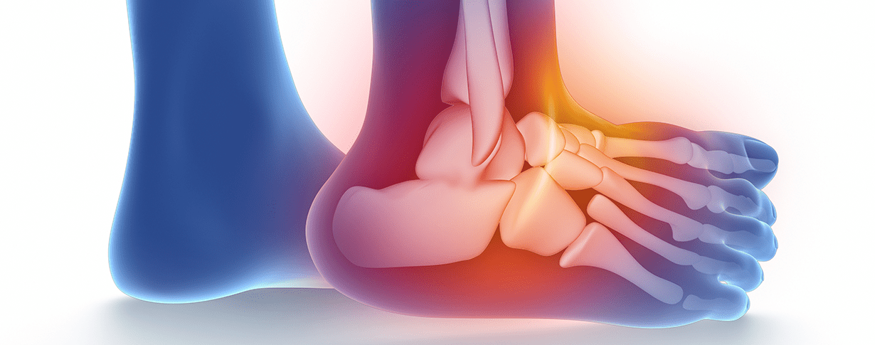
cartilage ; It is a solid, flexible, veinless and nerveless connective tissue consisting of collagen and elastic fibers, which is not as hard as bone and prevents bones from rubbing against each other by covering the joints.
It consists of 3 different types: hyaline cartilage, fibrous cartilage and elastic cartilage.
WHY DOES CARTRIDGE DAMAGE OCCUR?
1.Excess Weight: Since excess weight puts excessive load on the hips, knees and ankles, it causes faster wear and tear of the cartilages. If the problem that causes obesity is metabolic (such as diabetes), this problem should be eliminated. Therefore, lose weight and maintain the weight you lost.
2. Trauma: Cartilage damage also occurs due to injury. This injury may be minor but can grow over time with forces acting on the joint. Detected cartilage injuries should be treated immediately (PRP, microfracture, stem cell, etc.).
3. Joint Instability (Imbalance): Any ligament injury in or around the joint will cause excessive movement and friction of the joint, causing cartilage damage. When ligament injury is detected, it should be treated with methods such as ligament repair, ligament reconstruction (creating a new ligament), PRP etc. to protect the cartilage.
4.Nutrition: In order for the cartilage to be stronger and to repair the damage more easily, it especially needs some nutrients. In particular, adequate daily protein (meat, egg, pulses) intake, vitamin C (orange, lemon, strawberry) and vitamin E (corn, wheat, soy, almond and peanut) should be taken. These nutrients play an important role in the production and protection of cartilage.
5. Medications : It has been reported that cortisone administered into the joint or taken orally for a long time has negative effects on cartilage. For this reason, intra-articular cortisone is not recommended and long-term oral cortisone should be supplemented with cartilage-protecting drugs (glucosamine and chondrotin sulfate) and nutrients (protein C and vitamin E) when necessary.
6. Alignment Disorder: If there is congenital or subsequent curvature in the bones, this creates misalignment in the joints at the top and bottom of the bone. This misalignment also causes the cartilage to deteriorate as a result of overload. In order to preserve the cartilage, this misalignment must be properly corrected.
7.Avascular Necrosis (AVN): It is the melting of the cartilage as a result of anemia due to any obstruction of the blood vessels leading to the joint (trauma, cortisone use, metabolic diseases, alcohol). In the treatment, some drugs that open blood vessels should be used and if necessary, bone and cartilage should be revitalized by drilling holes (drilling).
8. Genetics: Lastly, age and genetic predisposition play a role. There is no procedure that can be done for genetic predisposition. With age, cartilage wears out and becomes more unstable.
NON-SURGERY TREATMENT:
The aim of the treatment in cartilage damage is to enable the patient to perform his/her daily life painlessly and fully perform joint functions. The decision to treat depends on the patient’s activity level, age, cause, and extent of damage, with or without surgery.
In cases where cartilage damage is mild, non-surgical treatment can be applied. This form of treatment is aimed at relieving the patient’s pain. This form of treatment includes: edema solvents (pain relievers), cartilage protectors (glucosamine & chondrotin and omega-3) intra-knee injections (hyaluronic acid, PRP), weight loss and Physical Therapy.
Hyaluronic Acid
Hyaluronic Acid (HA) is a viscoelastic substance and has a lubricating effect. This substance, which is specially prepared in the form of gel, is injected into the knee with the help of an injector. It can be done in 3 doses with one week intervals or a single dose with new generation drugs. This procedure can be repeated every 6 months, depending on the patient’s pain. HA effect does not start immediately. It is necessary to avoid excessive activity for a few days following the injection.
These injections have risks such as local swelling, allergies, infection and bleeding.
Glucosamine
Glucosamine-Chondrotin Sulfate is supportive treatment. Being in tablet form provides ease of use. Although there is no scientific data that this treatment prevents cartilage damage and calcification, it has been reported that pain is reduced in patients who apply this treatment.
PRP (Platelet Rich Plasma)
It is the separation of high-concentration blood cells called thrombocyte with the help of a special machine from whole blood taken from the patient. It is the injection of these separated cells into the joint with the help of a needle.
PRP is widely used, but its application form, amount and indications are not fully clarified. Generally, 2-8 ml is applied at 1-2-3 week intervals.
Diabetes, systemic disease, age and the drugs used by the patient generally affect the effectiveness of PRP.
Although it is widely used in the removal of intra-articular cartilage damage, muscle-beam diseases and ligament injuries, a standard treatment has not yet been obtained due to studies conducted with different methods and different patient groups.
Although many physicians have achieved very different results, the physician’s own experience is important in this practice.
.jpg)


.jpg)
Physical Therapy and Exercise:
Exercise is very beneficial as strengthening the muscles around the joint will reduce the load on the articular cartilage. In addition, some changes you will make in your daily life, for example; Using a cane, using an elevator instead of stairs, losing weight, swimming or walking in water will reduce pain. Resting and applying ice during periods of increased pain, and applying heat, stretching and stretching exercises when there is no pain are beneficial.
For this reason, age-appropriate sports should be done regularly.
Stem Cell (Lipogems)
It is the process of giving the stem cells obtained as a result of special processes from fat cells taken from the inner part of the abdomen or upper leg, to the joint with cartilage damage for structural support and repair. The procedure is performed in three stages (removal of adipose tissue, undergoing special procedures, giving it to the damaged tissue) in a single session under general or local anesthesia in operating room conditions and takes less than 1 hour.
It has been reported to give good results, especially in joints with stage 3-4 cartilage damage.